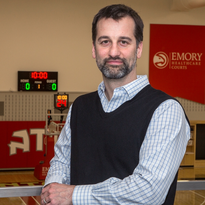
David Reiter, PhD
Jan. 31, 2018

When asked to explain his research, David Reiter’s short answer is this: “I use an incredibly versatile imaging tool to bridge voids in our ability to understand, detect, diagnose, and treat injury and disease.”
Sounds simple enough, but his work is quite complex. One aspect of Dr. Reiter’s research is focused on muscle, specifically the muscle’s dense network of tiny blood vessels, or microvasculature. He’s investigating ways MRI can measure the extent to which blood flow within the microvasculature affects muscle function and metabolism. This technical pursuit is important for understanding and treating impaired mobility due to metabolic diseases like diabetes, injury or other trauma, and even aging.
“We know older persons are not able to produce as much energy in their muscles, which affects their ability to run or walk fast,” says Reiter. “A lot of evidence suggests this decline in energy production arises from damage within the cell. The question is, to what extent is blood flow impaired and how does this impairment have an impact? Our preliminary research suggests a strong association between the delivery of blood to muscle cells—what we refer to as perfusion—and energetics in healthy aging. The more we understand the influence of microvascular supply and function in aging and disease, the better we can target interventions and therapies to improve health.”
Reiter is in the right place at the right time. He’s embedded in the Emory Orthopaedics and Spine Center at Executive Park which is connected to the new, state-of-the-art Emory Sports Medicine Complex, a joint venture with the Atlanta Hawks NBA team. There, he’s also involved in efforts to develop new methods for magnetic resonance imaging to diagnose connective tissue disorders, back disorders like discogenic disease, and problems with the articular cartilage of long bones.
“It’s very exciting, icing on the cake, to have a facility like the Emory Sports Medicine Complex,” he says. “It offers an extraordinary opportunity to develop advancements not just to benefit elite athletes but that also can be applicable to the general population.”
In many ways, Reiter says, elite athletes can present the ideal population for studying both optimal physiologic condition and musculoskeletal damage. “Advanced imaging can allow us to quantify training-induced metabolic and microvascular adaptations in athletes, for example. Alternatively, we can quantify and track changes in the cartilage of athletes’ joints after an acute injury. The same quantitative tools can tell us useful information in the study of diseases such as diabetes and osteoarthritis in the general population; it’s easy to see how we can benefit from comparisons between athletes and the general population.”
Reiter appreciates the opportunities for collaborative research that seems to abound at Emory. “From the beginning, since I first considered coming to Emory, I’ve been surprised and encouraged by the supportiveness of the leadership as well as by faculty at all levels. I think it’s a unique environment among top institutions and one that fosters community, sharing of ideas, and collaboration.”
A combination of “randomness and curiosity” led Reiter to a career in imaging sciences research. “I was drawn to engineering first because it’s quantitative and provides analytic tools for understanding and viewing the world,” he explains. “I also thought about a career in medicine because, as an athlete, I was fascinated by the human body, and particularly the musculoskeletal system.”
Engineering seemed to be a good way to combine his analytic acumen with his interest in human anatomy. At the University of California Davis, where he earned his PhD in mechanical engineering, he became part of the Biomedical Graduate Group, although he didn’t have a clear idea of what he would pursue. The chance to use small-bore MRI for a project changed everything.
“Here was this single instrument, a 7 Tesla Bruker Biospec, that allows you to noninvasively produce quantitative images of the body, mapping anything from tissue type, microstructure, mechanical strain, blood flow velocity, water diffusion properties, chemical composition, and enzymatic reaction rates. The versatility was mind-blowing.”
Now, as a member of the Emory Radiology Division of Research, Reiter is excited about the opportunities for mind-blowing on a much larger scale.
“It’s beautiful to be at an institution with such a clear articulation of its vision” he says of the department’s affinity for leading through innovation, a theme that resonates with his own approach to research. “Innovation in research doesn’t happen with the next obvious step in the same research process, but, rather, in developing new tools and new collaborative partnerships. I’ve always been driven by addressing important unmet needs. I’ve always had that spirit in what I’ve done. When your work is directed at identifying, understanding and addressing diseases that substantially impact human health, you are in a position to have a great impact.”


